

From Cell Type to Cell State in MS
Decades of genetic and immunological work settled which cell types matter in multiple sclerosis (MS). CD4 T cells and microglia sit at the top of that list, and no current paper spends effort arguing the point. What the single-cell literature is doing now is one level further down: not which cell type, but which state inside it, under what stimulus, in which part of which lesion. The shift is easy to miss because the vocabulary barely changes, yet it alters what a result can be used for. A cell type is a label you can sort on; a state is something a cell passes through, which means it can be caught early, or prevented. Three recent studies work at that level, and the third asks the awkward question of whether our laboratory models contain the states at all.
Which CD4 T Cell, Not Whether
Van Puijfelik and colleagues started from the observation that T helper 17.1 cells have been argued into a central role at MS onset largely on abundance, while almost nothing is known about their internal heterogeneity. They separated Th17.1 from Th17 and Th1 cells in healthy blood by spectral flow cytometry on chemokine receptor combinations, then followed brain-homing behaviour in paired blood and cerebrospinal fluid from untreated MS patients, and in blood drawn before and after six months of natalizumab. Purified subsets went to single-cell transcriptomics with matched stimulation assays. One cluster came out of all of it: Th17.1 cells carrying high CCR5, enriched in cerebrospinal fluid and selectively depleted from blood after natalizumab. The drug blocks entry to the brain, so a population that falls in blood under that treatment is a population that was heading there.
A Cell That Is Not Yet Cytotoxic
The transcriptional signature matched the phenotype and named a condition rather than an identity: receptive, and pre-cytotoxic. These cells carry CCR2, CD26, CXCR6 and granzyme K alongside high CCR5, and the killing machinery is not yet running. Interleukin 12 switched on STAT4 phosphorylation and raised the interleukin 18 receptor; interleukin 12 and 18 together raised granzyme B and perforin while granzyme K fell, and secretion of the cytolytic proteins appeared only when both cytokines were present. The cells that had gone through this carried GPR56 and CX3CR1. The authors also ran T cell receptor clonality and report a clean negative: none of the expanded clones matched any sequence in the reference database known to recognise a defined epitope, and the number of expanded clones did not differ with treatment. They state their own gap directly, that they have no direct evidence of these cells near active lesions, and note that gender, ancestry, race and ethnicity information for the participants was not available.
Two Microglial Shapes, Two Lesion Futures
Van den Bosch and colleagues took the same move into brain tissue. Lesion expansion drives disability while remyelination can recover function, and the question is what separates the two outcomes. Rather than compare lesions with healthy white matter, they compared lesions against each other, sorted by the shape of the microglia inside them: foamy, meaning lipid-laden, against ramified, meaning branched. Then they ran spatial transcriptomics at single-cell resolution on post-mortem human brain, so that every transcript keeps its coordinates. The result is that the molecular programmes are not mixed through the tissue but sorted by microglial state, which is a stronger claim than a difference in average expression and only available because the method keeps position.
What Sits Around Each Microglial State
The two neighbourhoods read very differently. Around ramified microglia the expression profiles concern myelin stability and axonal support, which the authors describe as an environment permissive for repair. Around foamy microglia sit immune activation, immunoglobulin production, complement activity, iron handling gone wrong, oligodendrocytes in an immune state, and demyelination itself. Calling these distinct immune-glial niches is the useful part, because a niche is addressable in a way a cell type is not: it has an inside and an outside, and a lesion can be asked which one it currently has. The same microglia, by lineage and by genetic risk, end up in two environments with opposite prospects. That is also why the comparison had to be lesion against lesion. Set against healthy white matter, both kinds of lesion would have returned the same answer, which is that a lesion differs from normal tissue, and the split that carries the prognosis would have been averaged away.
Whether the Models Contain the States
Aboelnour and colleagues asked the question the first two papers leave open. They integrated new and published single-cell datasets from two standard demyelination models, cuprizone and lysophosphatidylcholine, and set them against human MS data. Some of it carries across. Cuprizone produces a distinct stressed oligodendrocyte state marked by Cdkn1a and Nupr1 that resembles phenotypes seen in MS lesions, and both models converge during remyelination on an immune responsive oligodendrocyte state expressing Socs3, B2m and interferon response genes. Mouse microglia share a conserved activation programme, though lysophosphatidylcholine drives a stronger and longer one. Then the finding that matters for everything above: neither model captures the oligodendrocyte progenitor and microglial heterogeneity seen in MS. The states exist in patients and are missing from the models used to study them.
What a State Buys That a Type Does Not
Read together, the three mark a change in what counts as an answer. Knowing that CD4 T cells matter in MS has been true for decades and supports no intervention on its own; knowing that a CCR5 high Th17.1 cell sits one cytokine pair away from killing gives both a window and two molecules to act on. Knowing microglia matter supports nothing either; knowing that a lesion with foamy microglia carries a different molecular neighbourhood from one with ramified microglia turns a static label into a prognosis. The cost is that states are harder to work with than types. They are transient, they need position or stimulation to show themselves, and the third paper shows they may be absent from the standard models, which means the gap is not only between mouse and human but between what a model contains and what a patient has.
Disclaimer: This blog post is based on the cited studies and is intended for informational purposes only. It is not intended to provide medical advice. Please consult with a healthcare professional for any health concerns.
References:
van Puijfelik, F., Rip, J., van Hasselt, Y., et al., Smolders, J., & van Luijn, M. M. (2026). Refined single-cell profiling captures a CCR5 high CD4+ cytotoxic T-cell precursor in multiple sclerosis. EBioMedicine, 129, 106324. https://doi.org/10.1016/j.ebiom.2026.106324
van den Bosch, A. M. R., Khoo, J. H., Lu, Z., et al., Mulder, J., & Huitinga, I. (2026). Microglial states associate with lesion dynamics in multiple sclerosis. Cell Reports, 45(6), 117538. https://doi.org/10.1016/j.celrep.2026.117538
Aboelnour, E. L., Vanoverbeke, V. R., Maupin, E. A., Hatfield, M. M., & Adams, K. L. (2026). A comparative transcriptomic analysis of mouse demyelination models and multiple sclerosis lesions. Nature Communications, 17(1). https://doi.org/10.1038/s41467-026-72383-y
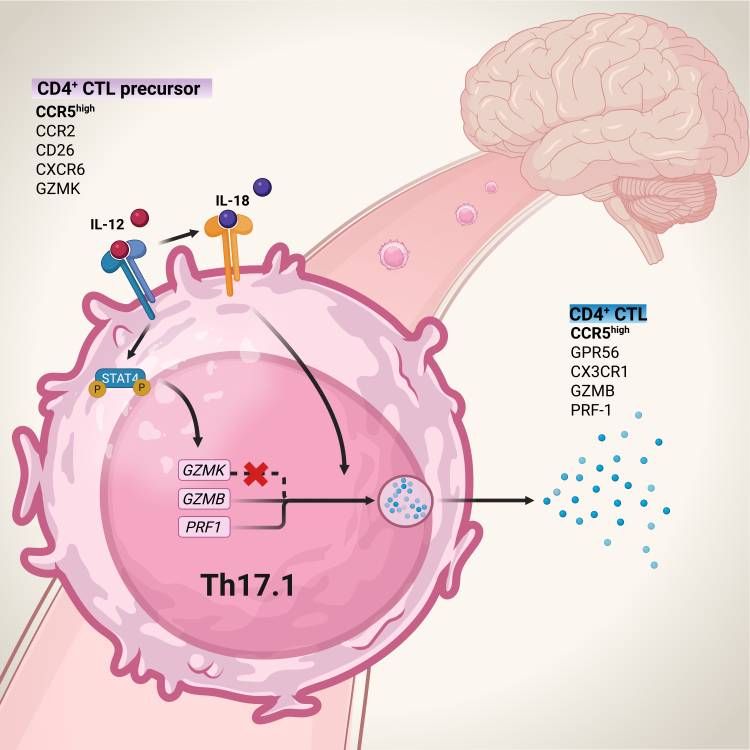
How the precursor becomes a killer: interleukin 12 drives STAT4 phosphorylation and interleukin 18 receptor expression on a CCR5 high Th17.1 cell, after which the two cytokines together raise granzyme B and perforin while granzyme K falls, and the cell takes on GPR56 and CX3CR1. (van Puijfelik, et. al. (2026), EBioMedicine 129, 106324, Figure 6, CC BY 4.0)