

Where MS Genetics Stops and Proteins Start

Most of what now gets called a multiple sclerosis (MS) protein comes from one of three kinds of study, and the word does different work in each. One measures proteins in the spinal fluid of living patients and reports which ones move with the disease. One never measures a protein in a patient at all, using genotype to stand in for lifelong protein level and asking whether that shifts MS risk. One takes brain tissue after death and reports which proteins sit inside the lesions that mark the fastest decline. Three papers published in the last year do one each, in large human cohorts, and the lists they produce overlap far less than the shared vocabulary suggests.
Twenty-Five Years of Spinal Fluid
Kosa and colleagues report a cohort assembled over 25.6 years at a single centre: 1,040 cerebrospinal fluid samples, of which 971 came from 394 people with MS and 69 from 44 healthy controls. Of 4,789 proteins measured, 3,714 passed a signal-to-noise threshold. The controls were used to subtract the effects of normal ageing and sex before anything else was tested, and what remains is attributed to disease. Most samples were drawn while patients were untreated, which is rare at this scale and is the reason the cohort can describe natural history rather than drug effects, and the full phenotypic range is present, with 162 relapsing-remitting, 151 secondary-progressive and 321 primary-progressive untreated samples. Genotype appears as a covariate rather than an explanation, with the intrathecal effects of HLA-DRB1*15:01 and *03 reported alongside those of demographic factors, body mass index and smoking.
What Moved With the Disease
1,668 proteins differed between patients and controls. Mapping each protein to the cell type that makes it showed plasmablast and plasma cell proteins raised by 90%, monocyte proteins by 56% and macrophage proteins by 52%, with oligodendrocyte proteins the only group reduced, by 73%. In the 110 patients who gave at least two untreated samples a median of four years apart, 910 proteins and 36 pathways changed significantly over time, 79% of them rising. Of 258 proteins tied to the number of contrast-enhancing lesions, the strongest included CXCL13, MMP9 and the heavy neurofilament chain. Sex differences survived the earlier correction: men carried higher levels of most proteins that rose with lesion counts, while women showed raised osteopontin. The authors note there is no external validation cohort, since no comparable untreated longitudinal collection exists, and call the mechanistic readings hypothesis-generating.
Genotype Standing In for a Protein
Belbasis and colleagues never drew spinal fluid. They took the lead genetic variant near each protein-coding gene that sets that protein's level in plasma, measured on the Olink and SomaScan platforms in UK Biobank and the deCODE study, and used it as an instrument in two-sample Mendelian randomisation against published genome-wide association results for Alzheimer's disease, Parkinson's disease, amyotrophic lateral sclerosis and MS. Because the variant is fixed at conception, an association between it and disease cannot run backwards from illness to protein, which is the whole point of the design. Across 2,738 proteins and four diseases they ran 13,377 tests. MS accounted for 3,492 of them and returned the largest yield of any of the four, with 377 associations at nominal significance, 64 surviving a 5% false discovery rate and 29 of those supported by colocalisation of the protein and disease signals at the same locus. Even after the significant results were removed, the remaining MS tests stayed inflated above the null by a factor of 1.41, the highest of the four diseases.
Which Proteins Carried a Direction
The MS list mixes familiar names with new ones. CD5, CD40, IL7R, STAT3, TNFRSF1A and thymidine phosphorylase were already tied to the disease, and alongside them came PARP1, parvalbumin and vascular endothelial growth factor B. Several point at innate immunity and at antiviral defence in particular: higher plasma levels of the poliovirus receptor came out protective, and the authors connect it to the control of viral microRNAs; tryptophanyl-tRNA synthetase and complement receptor 1 were also new. The design has limits the authors set out. The underlying association studies were built for disease risk and not progression, so these proteins are candidates for prediction or prevention rather than for treating established disease, and the two platforms together cover only part of the proteome.
Proteins Where the Damage Is
Van der Vliet and colleagues worked on tissue. From the Netherlands Brain Bank they took 110 fresh-frozen white matter samples from 28 donors with secondary progressive MS and 10 controls, sorted the lesions by histology, then split active and mixed lesions by whether their microglia were foamy or not. Across the full brain bank cohort, 250 donors and 8,708 lesions, a high proportion of foamy lesions went with a shorter time to the disability milestones, shorter disease duration and younger age at death, while remyelinated lesions ran the other way. Lipidomics across 712 lipid species, transcriptomics, proteomics and activity-based protein profiling on the same samples converged on lysosomal stress and disturbed lipid handling, with GPNMB, GRN and CHIT1 as hub genes of the module most tied to the foamy phenotype, and the module reproduced at protein level. Lesions called foamy held 52% foamy microglia on average, against under 1% in control and normal-appearing white matter, and they accumulated cholesterol esters and bismonoacylglycerolphosphates. Oxylipins in spinal fluid tracked the proportion of foamy lesions.
Three Senses of One Word
The three lists answer different questions. A protein on the first list is present in the compartment and moves with an outcome, which leaves open whether it causes anything or is released by dying tissue. A protein on the second carries a direction, because genotype fixes the exposure, but it was never measured in anyone with MS and the risk studies behind it say nothing about progression. A protein on the third sits in the tissue where decline is fastest, in people who have died, with no way back to the years when the lesion formed. Only one study here had genotype in a causal role, and it is the one furthest from the disease, working on blood from people who mostly do not have MS. The tissue and the causal claim have not yet been obtained from the same person.
Disclaimer: This blog post is based on the cited studies and is intended for informational purposes only. It is not intended to provide medical advice. Please consult with a healthcare professional for any health concerns.
References:
Kosa, P., Ashida, S., et al., Jennings, L., & Bielekova, B. (2025). Longitudinal proteomic profiling of cerebrospinal fluid in untreated multiple sclerosis defines evolving disease biology. Nature Communications, 16(1), 11012. https://doi.org/10.1038/s41467-025-65154-8
Belbasis, L., Morris, S., van Duijn, C., Bennett, D., & Walters, R. (2025). Mendelian randomization identifies proteins involved in neurodegenerative diseases. Brain, 148(7), 2412-2428. https://doi.org/10.1093/brain/awaf018
van der Vliet, D., Di, X., et al., Huitinga, I., & van der Stelt, M. (2026). Foamy microglia link oxylipins to disease progression in multiple sclerosis. Nature Neuroscience, 29(7), 1585-1598. https://doi.org/10.1038/s41593-026-02302-3
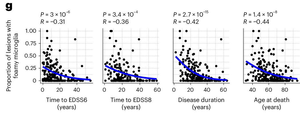
Across 250 brain donors and 8,708 lesions, the more of a donor's lesions contained foamy microglia, the sooner they reached each disability milestone and the younger they died. (van der Vliet, et. al. (2026), Nature Neuroscience 29(7), 1585-1598, Figure 1g, CC BY 4.0)