
What an MS Risk Allele Also Changes
A multiple sclerosis (MS) risk allele is named by a case-control table and then usually stays there, defined by the one thing it was tested against. Three papers published this year carry a genotype forward instead and measure it against something else: how much virus a person carries, what their T cells secrete, and what their brain looks like at autopsy. Two of the three never ask who has MS at all. Read together they show how much of what an allele appears to do depends on what the study chose to put on the other side of the equation, and they include one result that removes a mechanism the field has been assuming for years.
Genotype Against Viral Load
Kamitaki and colleagues went through whole-genome sequencing reads that did not match the human genome. Working across 490,401 blood samples in UK Biobank, 365,918 blood and 48,899 saliva samples in All of Us and 12,519 saliva samples in a third cohort, all sequenced at 32.5 to 42-fold coverage, they aligned the leftover reads against 31 common viruses and used the count as a measure of viral DNA load. Load varied with age, sex, season and time of day, and most of the viruses were more abundant in men. Common human variation at dozens of loci was associated with the load of seven of them: Epstein-Barr virus at 45 loci, human herpesvirus 7 at 37, and then herpesvirus 6B, Merkel cell polyomavirus and three anelloviruses. Smoking showed up as well, with viral read prevalence climbing steadily across pack-years. Ancestry mattered through a specific variant rather than in the abstract: the genotype that produces the malaria-protective Duffy-null blood type accounted for 28% of the difference in Epstein-Barr prevalence between people of African and European ancestry, and 45% of the difference for herpesvirus 7.
The MHC, for a Different Reason
For six of those seven viruses the strongest signal came from the MHC region, the same stretch of chromosome 6 that dominates MS susceptibility, and the region accounted for 56% of the heritability of Epstein-Barr virus load and 32% of herpesvirus 7. The alleles doing the work differed by virus. The top result for Epstein-Barr load was HLA-DRB1*04:04, where heterozygous carriers had viral reads 23.0% of the time against 13.6% in non-carriers, which the authors connect to the fact that the DRB1 molecule is the co-receptor the virus uses to enter cells. Outside the MHC the hits included ERAP1 and ERAP2, the peptidases that trim peptides for presentation. HLA-B*08:01 associated with load of one Epstein-Barr subtype and not the other, a host and virus genotype interaction.
A Null Result Worth Reading
Having found genetic instruments for viral load, the authors used 44 of them from outside the MHC in a Mendelian randomisation against MS, drawing on a meta-analysis of 7,907 cases and 1,474,810 controls. The estimate did not move from the null, with an MR-Egger p-value of 0.52, and the same analysis for Hodgkin's lymphoma came out positive across all four methods. People with MS in UK Biobank did carry more Epstein-Barr DNA than others, with a p-value of 0.005, so the association is real while the causal estimate is absent. The authors read this as lifetime viral load not adding further MS risk after infection, with the risk instead running through the immune response to particular viral peptides. The same instruments predicted Hodgkin's lymphoma before it appeared, at an odds ratio of 2.01 for carriers of detectable viral DNA.
A Risk Gene Read as a Switch
Pape and colleagues took one gene product rather than a genome. CD5 is a co-receptor of the T cell receptor complex, long described as a brake on signalling, and its gene is on the MS association list. Blocking CD5 on human memory CD4 T cells grown under Th17-polarising conditions cut production of interleukin 17A, interferon gamma and granulocyte-macrophage colony-stimulating factor without touching proliferation, and inhibiting casein kinase 2 cut two of the three while also slowing division. Both treatments lowered phosphorylation of STAT3, on different timescales. In a co-culture, Th17-polarised cells killed human neural cells, and blocking CD5 beforehand abolished that. Single-cell sequencing of spinal fluid from three patients and three controls found activation and homing genes raised in the CD5-positive CD4 T cells, a comparison that rests on six people. The larger human evidence came next: a 92-protein immunoassay run on 114 paired serum and spinal fluid samples, in which the samples with the highest CD5 also carried raised interleukin 17A, interferon gamma, CD137 and PD-L1, and in patients without relapse activity serum CD5 tracked neurofilament light, the standard marker of axonal injury.
Genotype Against What the Brain Looks Like
Lütje and colleagues went to 287 donors in the Netherlands Brain Bank, with an average of 21.8 tissue blocks and 35.4 lesions dissected per donor, and scored four features that belong to the donor rather than to any one lesion: perivascular cuffs, microglial nodules, mixed lesions with a broad rim of activated myeloid cells, and how much of the donor's lesion set had remyelinated. Cuffs were present in 25.5% of donors, nodules in 72.5%, broad rim lesions in 24.0%. Donors homozygous for rs3135388, which tags the main susceptibility allele HLA-DRB1*15:01, were more likely to show cuffs and nodules. Carriers of rs10191329 in the DYSF and ZNF638 locus, the severity variant, were enriched among donors with broad rim lesions, with cuffs at the edge of significance. Remyelination efficiency was associated with neither variant. Every donor homozygous for the severity variant had at least one of the four features, and only 29 of 204 donors had none of them.
What the Three Have in Common
In all three the genotype is the fixed quantity and everything else is the measurement, which is why the Nature result and the brain bank result sit together without contradicting each other. One says the amount of virus a person carries, itself partly set by HLA type, does not go on to change MS risk. The other says the HLA type is nonetheless visible in the brain decades later, as lymphocytes banked around vessels and clusters of microglia. Both can hold, because viral load and perivascular cuffs are different objects. What none of the three has is the same person measured twice. The brains come from people whose viral load was never counted, the biobank genomes come overwhelmingly from people who will never develop MS, and the T cell work used cells from donors with no pathology attached.
Disclaimer: This blog post is based on the cited studies and is intended for informational purposes only. It is not intended to provide medical advice. Please consult with a healthcare professional for any health concerns.
References:
Kamitaki, N., Tang, D., McCarroll, S. A., & Loh, P. R. (2026). The DNA virome varies with human genes and environments. Nature, 653(8116), 1099-1109. https://doi.org/10.1038/s41586-026-10288-y
Pape, K., Hanuscheck, N., et al., Bittner, S., & Zipp, F. (2026). Role of CD5 signalling for pro-inflammatory Th17 response in multiple sclerosis. Brain, 149(1), 123-133. https://doi.org/10.1093/brain/awaf268
Lütje, L., Chen, J. Q. A., Hamann, J., Smolders, J., Huitinga, I., & van den Bosch, A. M. R. (2026). Donor-specific pathological features associate with genetic background, lesion type distribution, and clinical heterogeneity in multiple sclerosis. Acta Neuropathologica, 151(1), 72. https://doi.org/10.1007/s00401-026-03040-3
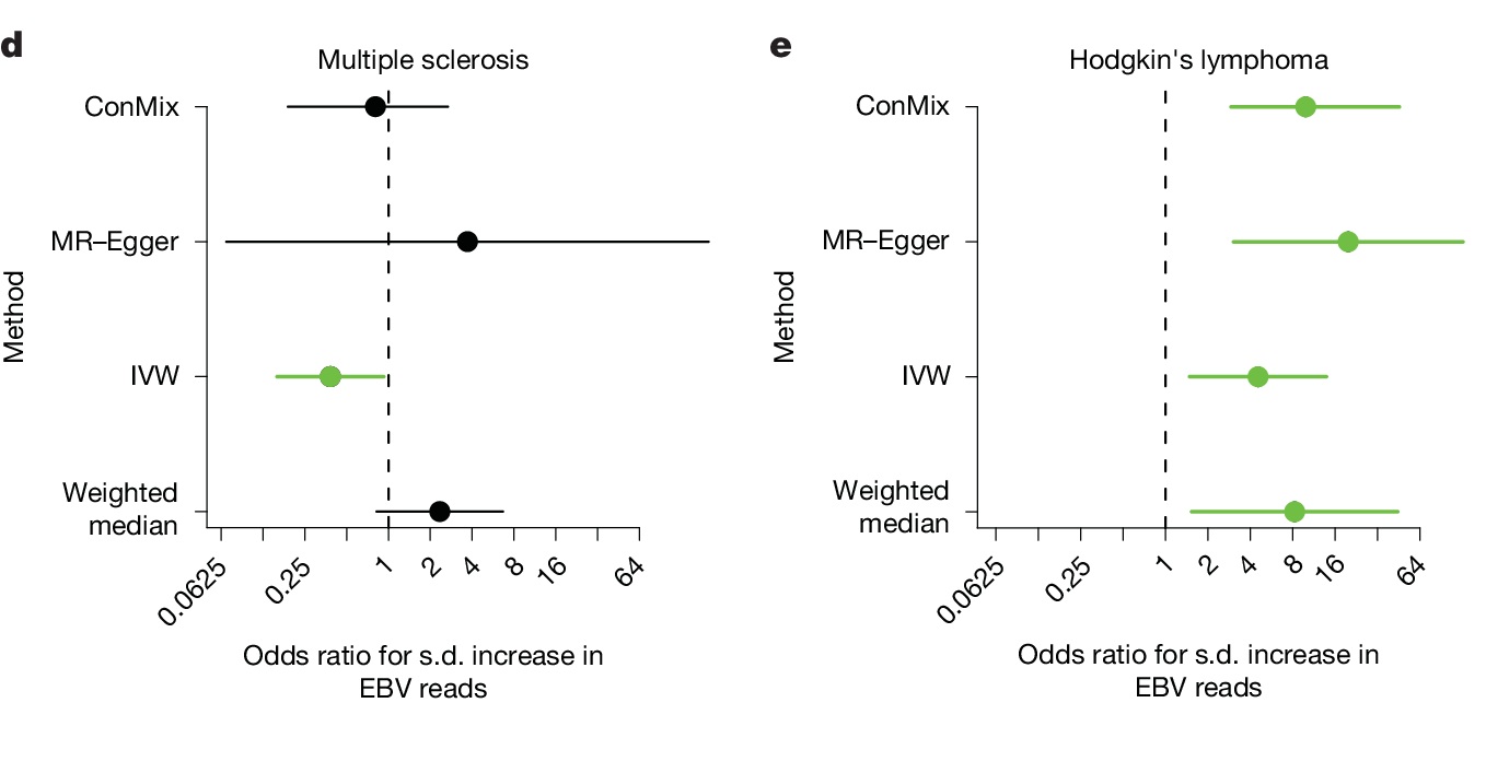
The same genetic instruments, two diseases. Every method crosses the null for multiple sclerosis and none does for Hodgkin's lymphoma. (Kamitaki, et. al. (2026), Nature 653(8116), 1099-1109, Figure 5d and 5e, CC BY 4.0)